

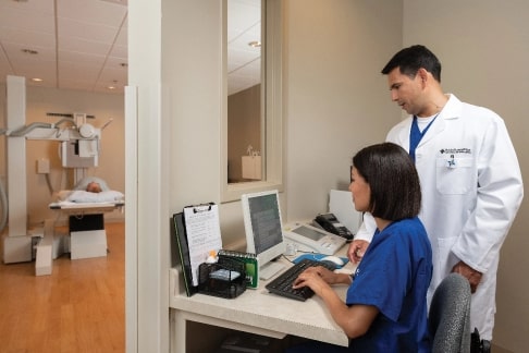
Imaging and radiology
Today, advanced imaging is the key to diagnose any health problem and start the healing process. In orthopedics especially, access to advanced diagnostic imaging is essential to an accurate diagnosis and effective treatment. We even use mobile imaging equipment in the operating room.
Have a doctor’s order already? Call us to schedule an Imaging appointment.
Here’s a closer look at the diagnostic imaging tools we use to help you heal.
CT (Computed Tomography)
A CT scan, also known as a CAT scan or Computed Tomography, is a special kind of X-ray that takes pictures of a cross-section of your body. CT scans may be used to find certain conditions that regular X-rays cannot find. CT scans also can be used to monitor progress during or after medical treatment for some conditions. The CT scanner is comprised of an X-ray tube that moves around your body. The CT scanner then sends signals to a computer.
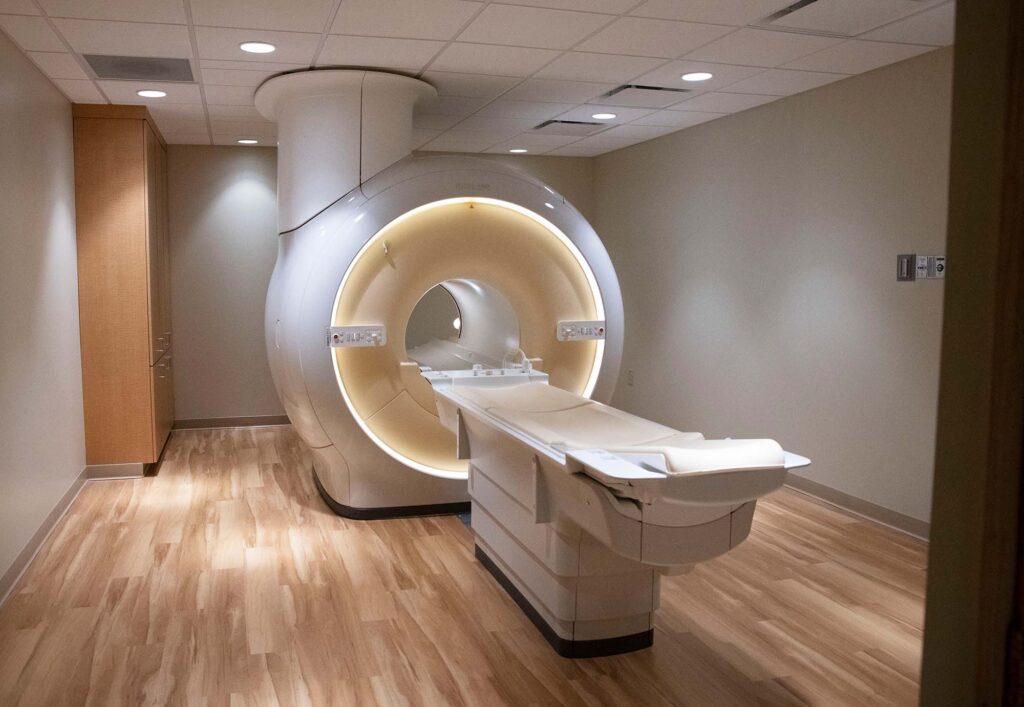
MRI (Magnetic Resonance Imaging)
Magnetic Resonance Imaging, or MRI, is a noninvasive, painless way to look inside the body at your organs and other body tissues. An MRI is a test that can find changes in your body, leading to early diagnosis and treatment of disease. MRI uses a large magnet, radio waves and a computer to create images of your organs and tissues. MRI does not use radiation, and there are no known side effects.
Unlike most hospitals, we read our MRI scans in-house instead of outsourcing to another facility or tele-radiology practice. This allows us to deliver your results much more quickly.
Ultrasound
Also known as sonography, an ultrasound uses high-frequency sound waves to capture live images inside your body. Ultrasound uses no radiation while visualizing muscles, tendons, ligaments, joints and even pinched nerves. It serves as a less expensive, but highly accurate, alternative to MRI. Doctors use ultrasounds to determine the source of pain, and can do so while the body part is in motion – rather than requiring you to remain completely still during a scan.
Digital Fluoroscopy & X-ray
A fluoroscopy is an examination of the tissues and deep structures of the body using X-ray imaging devices. One of these devices projects radiographic (X-ray) images in a movie-like sequence onto a screen monitor.
Arthrograms
Conventional arthrography is the X-ray examination of a joint that uses a special form of X-ray called fluoroscopy and a contrast material containing iodine. Arthrographic images help physicians evaluate alterations in structure and function of a joint and help to determine the possible need for treatment, including surgery or joint replacement. The procedure is most often used to identify abnormalities within the shoulder, wrist, hip, knee and ankle. The procedure is also used to help diagnose persistent, unexplained joint pain or discomfort.
Myelograms
A myelogram is an X-ray of the spaces in the spinal cord which contain various nerves. This test uses a special dye (contrast material), which mixes with the spinal fluid. Myelograms are useful for those who cannot have MRIs, including patients with pacemakers and cochlear implants.